A data-driven approach to study the mechanisms underlying altered reward processing in individuals with Parkinson’s Disease.
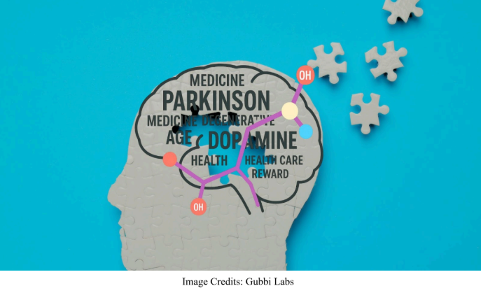
‘Parkinson’s disease’ (PD) is a neurodegenerative disorder mainly affecting the central nervous system. In 2020, more than 10 million people worldwide were living with Parkinson's disease, and 10% of affected individuals were in India. Patients with PD show symptoms including shaky limbs, muscle stiffness, and slow movements. While these movement-related symptoms are visibly apparent, individuals with PD also manifest other symptoms that are not related to movement (non-motor), such as lack of motivation or disability in experiencing pleasure, attributed to a lack of dopamine hormone. Commonly known as the ‘feel-good’ hormone, dopamine is generally produced when performing a pleasurable task or receiving a reward.
The ability to appreciate pleasure and rewards is a fundamental aspect of human well-being and quality of life. Dopamine provides a pleasurable sensation, motivating one to seek more of the sensation or repeat the rewarding behaviour. Lack of dopamine in individuals with PD leads to altered brain activity and impaired reward processing – the brain's ability to recognise, value, and respond to rewarding stimuli.
In their recent study, researchers from the Human Motor Neurophysiology and Neuromodulation Lab, Department of Biosciences and Bioengineering at the Indian Institute of Technology Bombay (IIT Bombay) used brain signals to investigate reward processing in individuals with PD.
“In PD, motor symptoms like stiffness and tremors are typically the first noticeable signs, while the non-motor symptoms, including cognitive and emotional changes, often emerge years earlier. Some patients experience cognitive or emotional changes early on, while others may develop them much later, making it difficult to establish a consistent sequence of symptom onset,” remarks Prof. Nivethida, who led the study.
Brain cells (neurons) communicate with each other through electrical signals. Electroencephalography (EEG) measures the electrical activity in the brain using small metal discs placed on the scalp. When an individual performs a certain task, EEG detects changes in the electrical activity pattern of the brain regions that are involved in the task. In this study, the researchers analysed EEG data recorded from 28 PD patients and 28 healthy individuals while they performed a reward-based learning task. EEG was recorded from PD patients both before (OFF condition - 15 hours after their last dose) and after taking dopamine medication (ON condition). Comparing these conditions allowed researchers to understand the influence of dopamine medication on reward processing.
The researchers then used three different analysis methods to understand the brain activity linked to reward processing. First, they used Event-related Potential (ERP) analysis, which computes the brain's response to a specific sensory, cognitive, or motor event. They found that 250-500 milliseconds after receiving a reward, the ERP from the front part of the brain generally shows a positive response, a phenomenon called reward positivity (a positive waveform response on EEG linked to a reward). Reward positivity is essential for cognitive processes like attention, learning, and emotional responses.
Next, the researchers used time-frequency analysis, which identifies the periodicity or rhythm in the brain activity. They identified slower brainwaves or theta waves with frequencies between 5-7 Hz and faster brainwaves or gamma waves with frequencies between 30-55 Hz, each associated with distinct cognitive states or brain activity. Theta waves are linked to reward processing and creativity, while gamma waves are associated with decision-making and problem-solving. Finally, they measured the level of synchronisation between the theta and gamma waves using a method called phase-amplitude coupling (PAC), which is thought to underlie communication between brain regions. Theta-gamma coupling or synchronisation is crucial for cognitive functions such as reward processing and goal-oriented behaviour.
The results of this study show that reward positivity, as seen in the ERPs, was weaker in PD patients, indicating that their brains do not process rewards effectively. Further, dopamine medication failed to restore the reward positivity. “Normally, the brain releases dopamine as short bursts following a reward, but in PD, these bursts are weaker. Although dopamine medication replenishes the dopamine levels in the brain, it does not produce burst-like signals that mimic the natural process. This could be the reason why dopamine is able to improve motor symptoms but not cognitive functions like reward processing. Hence, adjunct treatment strategies may be required to restore cognitive impairments in PD,” adds Prof. Nivethida.
The results of the time-frequency analysis reveal that while healthy people show reward processing wave activity after receiving a reward, PD patients show weaker corresponding signals in both ON and OFF conditions. This indicates that PD individuals have less reward sensitivity, even with dopamine medication. “These results also suggest that reward processing mediated by theta activity may not be driven purely by dopaminergic mechanisms and that the role of other chemicals in the brain (neurotransmitters) should not be ignored,” says Prof. Nivethida.
The results of PAC analysis indicate that PD patients have weaker theta-gamma synchronisation, resulting in poor communication between brain regions that process reward information and use it for learning goal-directed behaviour. This could be the reason behind the lack of motivation and impaired decision-making in PD patients. Dopamine medication was able to partially restore theta-gamma synchronisation. This finding highlights theta-gamma coupling as a potential biomarker to identify the impairment of reward mechanisms in PD.
“Reward processing deficits are not only reported in PD. They are also observed in other neuropsychiatric conditions such as depression, schizophrenia, and other movement disorders. This overlap complicates its use as a specific early biomarker for PD without additional supporting evidence,” suggests Prof. Nivethida.
Additionally, the researchers report that patients showed higher levels of faster gamma activity in the back part of the brain than healthy individuals and that those with longer disease duration had higher gamma activity. It indicates that this gamma activity pattern could be linked to the disease process in itself and not linked to reward processing. Overall, the study highlights the role of disrupted brain activity patterns in PD patients and how they impact specific cognitive functions such as reward processing.
The IIT Bombay study clearly highlights the connection between atypical brain activity and the impaired ability of PD patients to appreciate rewards, in addition to delving into understanding reward processing in our daily lives. It offers valuable insights into the complex neural mechanisms underlying PD and emphasises the need for adjunct treatment approaches, such as noninvasive brain stimulation, for improving non-motor symptoms in individuals with PD. Notably, it sheds light on how maintaining optimal levels of motivation through reward processing contributes to the quality of our lives.
Prof. Nivethida Thirugnanasambandam,Department of Biosciences and Bioengineering, Indian Institute of Technology Bombay, Powai, Mumbai 400076, India.