Spinning disc confocal microscope facility
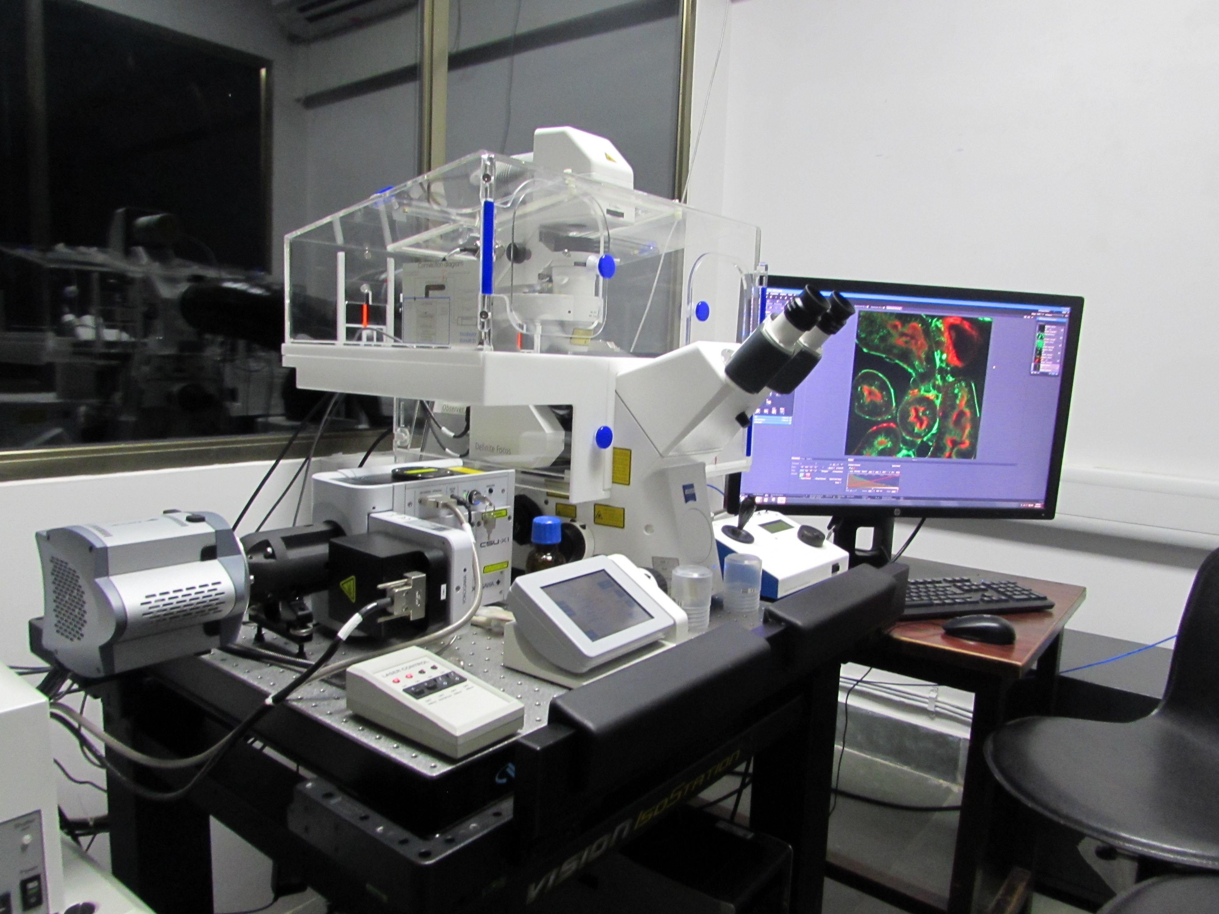
Spinning-disk confocal microscope facility is suitable for capturing fast changing phenomena, such as, imaging swimming bacteria, etc. Most powerful tools for live cell imaging.
Make and Model
Yokogawa Electric Corporation, CSU-X1
Available mode for use
-
Single colour and two-colour imaging and brightfield imaging
-
Z-stacks
-
Time series (with or without Z-stack)
Specifications/Features
-
Microscope and spinning disc: Zeiss observer Z1 inverted motorized and computer-controlled fluorescence microscope fitted with high speed microlens-enchanced Nipkow spinning disc
-
Objectives: 10X/0.45 NA (air), 20 X/0.8 NA (air), 40 X/1.2 NA (air), 40 X/1.3 NA (oil), 63 X/1.4 NA (oil) & 100 X 1.4 NA (oil). DIC imaging is possible with all the objectives
-
Lasers: 488nm and 561 nm solid state lasers as excitation sources
-
Image acquisition at 50 fps at full resolution.
-
Suitable for observing fast dynamic cell processes
-
Two colour (alexa fluor 488 and rhodamine)fluorescence imaging possible.
Application
-
Single colour and two-colour Imaging and brightfield imaging.
-
Z- Stacks
-
Time series (with or without Z-stack)
-
Fast dynamic processes
-
Multi-position imaging
Facility in-charge
Contact Email
sdconfocal[at] iitb[dot] ac[dot] inContact no.
022-2159 6746Location
Central Instrumentation Room, Ground floor
Biosciences & Bioengineering Department.
I.I.T. Bombay,Powai,
Mumbai - 400 076
Other contact person(s)
- Pradip Shinde
- Santosh Panigrahi
Facility Management Member(s)
(w.e.f. )
Prof. S PatankarProf. Santanu K. Ghosh
Prof. Swapnil Shinde
Prof. Sushil Kumar
Prof. Abhijit Majumder
Prof. Sandip Kar
Registration Link:
Information-IIT Bombay users
Internal user's information linkInformation-External users
External user's information link265.58 KB- Microscope and spinning disc: Zeiss Observer Z1 inverted motorized and computer-controlled fluorescence microscope fitted with high speed microlens-enchanced Nipkow spinning disc (Yokogawa CSU-X1 automated model). Peltier-cooled monochrome EMCCD camera with 56 fps frame rate at full resolution (512x512) for imaging in confocal mode. 1.4 MP monochrome camera with one-stage Peltier cooling mechanism for imaging in non-confocal (widefield) mode. Temperature (from 3oC to 45oC) and CO2-controlled incubation stage. Stage insert available for glass slides and 35mm Petri dishes. Proprietary Definite Focus from Zeiss with NIR LED Hardware Focus to eliminate drift during long-term imaging. Zeiss ZEN Blue image acquisition software
- Objectives: 10X/0.45 NA (air), 20X/0.8 NA (air), 40X/1.2 NA (air), 40X/1.3 NA (oil), 63X/1.4 NA (oil). DIC imaging is possible with all the objectives.
- Lasers: 488nm and 561nm solid state lasers as excitation sources.
- Filters for widefield (non-confocal) imaging: alexa fluor 488, rhodamine, cy5 and DAPI are currently mounted
- Image acquisition at 50fps at full resolution.
- Suitable for observing fast dynamic cell processes
- Two colour (alexa fluor 488 and rhodamine) fluorescence imaging possible. DAPI imaging is not available in the confocal mode.
- Live cell imaging possible
There is a trade-off between image acquisition at high-resolution and at high speed with conventional scanning probe confocal microscopy. This is because each point in the sample plane is scannedin a sequential manner to obtain a 2D image. In a spinning-disk confocal microscope, multiple points within the sample are excited simultaneously. The image is recorded using an array detector like a high-speed sensitive EMCCD camera instead of a point detector (e.g. PMT). This system is suitable for capturing fast changing phenomena, such as, imaging swimming bacteria, etc.
The Yokogawa CSU-X1 spinning disc has a combination of upper and lower disks rotated by a motor. The laser light is first defocused to expand to a larger spot size. This larger laser beam spot is then translated into ~20000 small focused laser beam spots by the upper microlens array disk. These laser beam spots then pass through a dichroic mirror and are perfectly aligned to pass through corresponding pinholes on the lower pinhole array disk. The laser spots are then focused by the objective lens onto the sample. Fluorescent light from the specimen returns along the same path through the objective lens and pinholes, is reflected by a dichroic mirror, and is focusedat a camera or eyepiece. Thus, the light beams can illuminate the entire observation area of the specimen and forms a confocal optical slice at the camera or eyepiece
Figure 1. The basic configuration of the Yokogawa CSU- X1 spinning disc is shown.
Date | Presentation File | Presentation by (Prof.) | Department | |
---|---|---|---|---|
04-12-2017 | View Presentation5.22 MB | Prof. Debjani Paul | Biosciences and Bioengineering | |
28-02-2019 | View Presentation15.32 MB | Prof. Debjani Paul | Biosciences and Bioengineering |
This FAQ deals with the operational aspects of the facility. If you would like to suggest a question, do feel free to drop an email to <debjani [dot] paul [at] iitb [dot] ac [dot] in>
1. I need to do simple slide imaging. Which confocal microscope should I use?
You could use either as long as you image up to two fluorophores (red and green). If you want to image DAPI, you have to use the scanning probe confocal microscope. There is no laser for DAPI excitation in the spinning disc system. If you want to image more than two fluorophores, you need to use the scanning probe confocal microscope.
2. What consumable items should I bring with me? What items will be provided at the facility?
The facility will only provide the immersion oil for the objectives and the lens cleaning tissues. Everything else that you may need during imaging (e.g. gloves, pipettes, tips, regular tissue rolls, aluminium foil to cover NDD, etc.) you will have to bring yourself. If you are in doubt, please speak to the operators or one of the conveners in advance.
3. I need to use the confocal microscope. Do I need to train as a TA?
If your usage is infrequent (less than twice per month), one of the operators or the existing TAs can do the imaging for you. If your research project heavily depends on the use of the confocal facility, it would be better if you trained as a TA. Do remember, training as a TA comes with certain duties, such as, imaging other people’s samples.
4. What does being a TA involve?
The job of a TA is to help us run the facility smoothly and image other people’s samples. You will have a do a minimum of 6 hours of TA duty per week just like the TAs allotted to other central facility equipment. This is non-negotiable. If you are a first year PhD student with loads of coursework, we suggest that you come back after a year. The upside is that you will get really proficient in using a stateof-the-art confocal microscope. You will also be able to book slots during ‘off’ hours (between 6pm and 9am) to run samples for yourself or your research group. On the whole it should be a very useful learning experience for you.
5. I think I need to train as a TA. What should I do?
The first thing you should do is to check with your advisor on whether both of you agree with the time commitment. If you are a non-BSBE student, you need to contact the TA coordinator of your department to see if you could be assigned as a TA in the central facility. If the answer to both questions is ‘yes’, send an email to the convener of the microscope where you want to train. We will take over from there.
6. I booked a slot, but my sample is not ready. What should I do?
This can happen once in a while, so don’t worry. Send an email to <confocal [at] bio [dot] iitb [dot] ac [dot] in> and call the operator on his mobile phone as soon as you realize that you cannot make it to your slot. This is a matter of courtesy to ensure that other people can use your slot. If this happens too many times, clearly you are not planning your experiments very well and we will take a strict view of it.
7. I need to do live cell imaging. Which microscope should I go for?
If you are imaging swimming bacteria, sperm cells, etc. you should definitely choose the spinning disc system. If your live cell imaging involves three or more fluorophores (unlikely though!), you have to choose the scanning probe confocal system. If your sample is tagged with a single fluorophore (green or red) and you want to image for an hour or so, you can choose either. If you want to do longer experiments (e.g. exploring the motility of a mammalian cell), we will assign you the spinning disc system unless there is a compelling reason to use the other microscope. Such experiments should be scheduled at night (after 8pm). If they need to run longer than 12 hours, you should plan to do these experiments over the weekend. In case of initial overnight operation, you (or the TA) will need to check on your sample every couple of hours.
8. I need to book more than one consecutive slot. Can I get it?
Sure, if you can justify why. Having ~20 odd samples to image at one go is not a good enough reason. We need to be fair to every user while assigning slots. We will give you as many slots as you need to image all your samples, but they will be distributed over several days.
9. I don’t have anyfluorophore in my sample. Can I still use the confocal system?
That depends. If you are doing live cell imaging (with CO2 and temperature) in brightfield/DIC mode for a long enough time, you certainly can use the system, as there is currently no other microscope in IITB that has this facility. Just remember that it won’t be confocal imaging, i.e. you won’t be blocking the out-ofplane light. In such a case, the Definite Focus feature will be very useful to you to ensure that at least one of the Z-stack images remains in focus throughout.
10. Can I request a particular TA to image my sample?
No. All TAs have done the same training and it should not matter who images your sample. The conveners have framed this policy to ensure that no single lab/TA monopolizes the use of the facility. If you have any apprehensions about any TA, feel free bring it to the notice of the convener immediately.
- MEHENDALE, N., SHARMA, O., PANDEY, S., & PAUL, D. (2018). “Clogging-free continuous operation with whole blood in a radial pillar device (RAPID)”. Biomedical Microdevices :BioMEMS and Biomedical Nanotechnology. 20, 1-1
- SURVE MV, BHUTDA S, DATEY A, ANIL A, RAWAT S, PUSHPAKARAN A, SINGH D, KIM KS, CHAKRAVORTTY D, & BANERJEE A. (2018). “Heterogeneity in pneumolysin expression governs the fate of Streptococcus pneumoniae during blood-brain barrier trafficking”. PLoSPathogens. 14.
- VENUGOPAL B, MOGHA P, DHAWAN J, & MAJUMDER A. (2018). “Cell density overrides the effect of substrate stiffness on human mesenchymal stem cells' morphology and proliferation”. Biomaterials Science. 6, 1109-1119.
- SHARMA H, JOHN K, GADDAM A, NAVALKAR A, MAJI SK, & AGRAWAL A. (2018).” A magnet-actuated biomimetic device for isolating biological entities in microwells”. Scientific Reports. 8.
- SRIVASTAVA S, & PANDA D. (2018). “A centrosomal protein STARD9 promotes microtubule stability and regulates spindle microtubule dynamics”. Cell Cycle (Georgetown, Tex.).
- Subhadeep Das, Rakesh Kumar, Narendra Nath Jha, and Samir K. Maji* , “Controlled Exposure of Bioactive Growth Factor in 3D Amyloid Hydrogel for Stem Cells Differentiation”, Adv. Healthcare Materials, 2017.
- Shalini Srivastava1and Dulal Panda1*, “A centrosomal protein FOR20 regulates microtubule assembly dynamics and plays a role in cell migration”, Biochemical Journal , 201
- “MEME Aptasensor for Label-free Detection of cancer cells “ Madhuri Vinchurkar, Mamta Ashwin, Anjali Joshi, Archana Singh, Prakriti Tayalia, V. Ramgopal Rao,: a:Department of Electrical Engineering, Indian Institute of Technology Bombay, Mumbai, India b:School of Biosciences and Bioengineering, Indian Institute of Technology Bombay, Mumbai, India c: Indian Institute of Technology Delhi, Delhi, India
- Lakshmi Kavitha Sthanam a, Amlan Barai a, Anuj Rastogi a, Vijay K. Mistaria, Ana Maria b,RahulKauthaleb, Madhumanjiri Gatne b, Shamik Sen a, * , “Biophysical regulation of mouse embryonic stem cell fate and genomicintegrity by feeder derived matrices”, 2016.
2021-2022:
1. Venkatramani, A., S. Mukherjee, A. Kumari, and D. Panda, Shikonin impedes phase separation and aggregation of tau and protects SH-SY5Y cells from the toxic effects of tau oligomers. Int J Biol Macromol, 2022. 204: p. 19-33.
2. Siddiquie, R.Y., A. Gaddam, A. Agrawal, S.S. Dimov, and S.S. Joshi, Anti-Biofouling Properties of Femtosecond Laser-Induced Submicron Topographies on Elastomeric Surfaces. Langmuir, 2020. 36(19): p. 5349-5358.
3. Pradhan, A., S. Mishra, A. Surolia, and D. Panda, C1 Inhibits Liquid-Liquid Phase Separation and Oligomerization of Tau and Protects Neuroblastoma Cells against Toxic Tau Oligomers. ACS Chem Neurosci, 2021. 12(11): p. 1989-2002.
4. Pradhan, A., S. Mishra, S.M. Basu, A. Surolia, J. Giri, R. Srivastava, and D. Panda, Targeted nanoformulation of C1 inhibits the growth of KB spheroids and cancer stem cell-enriched MCF-7 mammospheres. Colloids Surf B Biointerfaces, 2021. 202: p. 111702.
5. Mukherjee, S. and D. Panda, Contrasting Effects of Ferric and Ferrous Ions on Oligomerization and Droplet Formation of Tau: Implications in Tauopathies and Neurodegeneration. ACS Chem Neurosci, 2021. 12(23): p. 4393-4405.
6. Kumari, A., O. Shriwas, S. Sisodiya, M.K. Santra, S.K. Guchhait, R. Dash, and D. Panda, Microtubule-targeting agents impair kinesin-2-dependent nuclear transport of beta-catenin: Evidence of inhibition of Wnt/beta-catenin signaling as an important antitumor mechanism of microtubule- targeting agents. FASEB J, 2021. 35(4): p. e21539.
7. Sane, A., S. Sridhar, K. Sanyal, and S.K. Ghosh, Shugoshin ensures maintenance of the spindle assembly checkpoint response and efficient spindle disassembly. Mol Microbiol, 2021. 116(4): p. 1079-1098.
8. Saha, R., S. Patkar, D. Maniar, M.M. Pillai, and P. Tayalia, A bilayered skin substitute developed using an eggshell membrane crosslinked gelatin-chitosan cryogel. Biomater Sci, 2021. 9(23): p.7921-7933.
9. Patwardhan, S., P. Mahadik, O. Shetty, and S. Sen, ECM stiffness-tuned exosomes drive breast cancer motility through thrombospondin-1. Biomaterials, 2021. 279: p. 121185.
10. Malankar, G.S., A. Sakunthala, A. Navalkar, S.K. Maji, S. Raju, and S.T. Manjare, Organoselenium- based BOPHY as a sensor for detection of hypochlorous acid in mammalian cells. Anal Chim Acta, 2021. 1150: p. 338205.
Minutes of the user meeting held on 28th Sept 2016
Location: BSBE seminar room A
A joint meeting of the users (students, project staff and faculty members) of the laser scanning probe and spinning disc confocal systems was arranged. The meeting was attended by 23 student/project staff users, 5 faculty members (including the faculty members in-charge Santanu Ghosh and Debjani Paul) and the two operators.
A. Technical issues:
It was decided to request the Zeiss technical support team to come down and sort out the following technical problems.
1. A 405 nm laser source is urgently needed for DAPI imaging in the spinning disc (SD) confocal microscope so as to overcome the underuse of the system during working hours. We will send a request to IRCC with usage statistics and quotations.
2. Many users require DIC images along with fluorescence. Currently it is not clear how both can be done together in the spinning disc system. The engineers need to be contacted to check (i) whether this is possible, and (ii) what additional accessories are required to do this.
3. Several users reported a strong Z-drifting in the SD system, such that the entire sample goes out of focus within an hour. It happens during Z-stack acquisition at a single location, as well as while imaging at the multiple locations. This problem has not been seen in the LSM system. We need a clarification from Zeiss whether the definite focus systems are working differently in LSM and SD microscopes. We also need the Zeiss engineers to solve this at the earliest.
4. The ZEN Blue software in the SD system continues to crash repeatedly. This problem was reported at the last user/TA meeting as well.
Action items:
a) Faculty members in-charge and operators to follow up with Zeiss for technical support.
b) Operators to generate the usage statistics for users who request DAPI imaging. Faculty members in-charge to submit the request for DAPI laser to IRCC with quotes and usage statistics.
B. Slot booking options on Drona after working hours:
5. The slot booking maintained on the Google calendar and Drona need to match. Currently there is no way to book slots after 6pm on the Drona calendar. We need to send a request to IRCC software team to allow booking of slots between 6pm and 9am.
Action item: Faculty-in-charges to send requestto IRCC software team.
C. Policy for long duration imaging in the SD system:
6. Many users book the SD system only for live cell imaging for long durations. It is convenient to do these experiments overnight. Since the last TA duty slot gets over by 6pm, it was decided that these slots can start from evening 6:00 pm itself and the sample can be taken out at 9 am the next day.
7. Until the Z-drift problem is sorted and the DAPI laser is bought, the SD system is not used heavily during the day. Therefore, it was decided that multiple slots to a single user or slots for long duration imaging can be allotted during the day. Tuesdays and Thursdays are to be keptexclusively for long duration imaging, in addition to after 6pm and on weekends.
Action item: Disseminate the information to the confocal user group by email.
D. Additional TA duty:
8. Since there is a high usage of the spinning disc system after hours and also during the weekends, it was decided to allot TA duty for an additional slot after 6pm (most likely 6- 8pm) and also during the day on Saturday and Sunday (2 hour each). This is required for the users who are not TAs to be able to check on their samples during these long imaging slots.
9. There is also a requirement to train more TAs to manage the after-hour and weekend slots. A request will be sent around the institute in November/December for training of another batch of TAs. So far, 8 TAs have been trained on each microscope and the two operators also help in imaging.
10. Cleaning of the cell culture incubator and laminar flow hood, and emptying of the dehumidifier at least once a day on Saturdays, Sundays and holidays are to be included in TA duty. These duties will be carried out by all the TAs in rotation once a week.
11. Vaccuuming of the confocal rooms is to be done to minimize the dust related problems. A request to procure a vaccuum cleaner exclusively for use in the BSBE ground floor central facility will be made.
Action items:
a) Disseminate the information to the TA group by email
b) Operators to draw up new duty chart for TAs starting next week.
E. Slot cancellation policy:
12. The users are requested to inform of any cancellation in advance by any one of the three ways: (i) sending an email to the confocal users Google group, or (ii) sending a text message on the Whatsapp group of the users, or (iii) sending a text message to either the TA or operators in-charge. The TA/operator will then immediately send out an email to the user group informing of the available slot. Only verbal intimation for slot cancellation will not be accepted.
13. The first person to respond to the slot cancellation message by any of the above ways gets to use the cancelled slot. Verbal requests for slot booking will not be accepted.
14. If a user does not show up for 15 minutes into the slot without informing the TA or the operators, then that user will not be given a confocal slot for the next 1 month as a disciplinary action.
Action item:
a) Disseminate the information to the confocal user group by email.
b) Operators to inform new users about the cancellation policy when assigning slots.
F. User groups:
15.The confocal user Google group has recently been formed. It includes all users and their advisors. Other than reporting operational issues, this group will also be used by users to report any technical problem at the earliest.
16. A Whatsapp group of the users will be made by the operators for reporting slot cancellation only.
Action items:
a. Disseminate the information to the confocal user group by email.
b. Operators to make a Whatsapp group of the user base as a back-up contact strategy.
G. Information given to the users:
17. Both internal and external users will eventually be charged for using the two confocal systems. The modality for charging is being worked out by the facility management group in consultation with IRCC. The users will be informed as soon as a decision is reached.
18. A workshop on the usage of the two confocals will be organized to increase the user base within the institute.
19. We will organize a similar lecture and demonstration workshop to increase external (outside IIT) usage.
20. FLIM, FCS and FCCS training will be scheduled in November.
21. It was decided to showcase as artwork some of the good images obtained using the facility. A competition will be organized to select the images.
22. Users are requested to acknowledge the IRCC Central Facility whenever they use these images in any publication, conferences, etc.
Action item: Disseminate the information to the confocal user group by email.
- Currently we can image fixed samples sealed between a glass slide and a cover slip. Do not bring samples without sealing them with a cover slip.
- 35 mm diameter petri dishes. Please use specially available imaging petridishes with cover slip bottoms if you wish to use oil immersion objectives.
- Only online registration through the IRCC webpage will be accepted. If you need to cancel your slot, send an email immediately to with an explanation.
- Slots will be provided on a first-come first-served basis.
- The slots are from 9am - 11am, 11am - 1pm, 2pm - 4pm, 4pm - 6pm. You can request two consecutive slots only once in a week. If your experiment needs more time (e.g. long time live cell imaging, etc.), please drop an email to sdconfocal [at] iitb [dot] ac [dot] in and CC Prof. Debjani Paul debjani [dot] paul [at] iitb [dot] ac [dot] in so that we can deal with your specific requirement.
- USB drives are strictly not allowed for copying data to minimize virus-related issues. You need to bring a new blank CD to transfer your data. All data must be transferred within 7 days of imaging. Without exception.
- Users must be available throughout the imaging.
- Please mention what fluorophores you have used in your sample (excitation/emission spectra) when you make a request.
- Register online through the IRCC webpage.
- After the slotbooking request is accepted, please contact the operator (Pradip Shinde or Santosh Panigrahi at 4770) to discuss the details of your experiment.