This patent describes a scanless optical tomographic system, which simultaneously uses more than one optical line sources to perform a three-dimensional reconstruction of the optical and dynamic properties of turbid media.
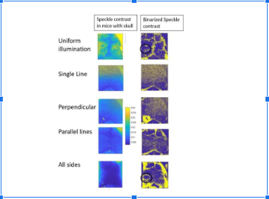
Figure 1. The blood flow in mice brain with their skull intact imaged by using this system
Traditional optical tomography systems require scanning the sample using a point source or a single line source to gather sufficient data for three-dimensional reconstruction. This scanning process involves complex instrumentation, such as electromechanical devices like Galvo mirrors, and reduces the system's temporal resolution. Conventional methods using point sources or line sources with scanning also suffer from limited signal-to-noise ratio and dynamic range.
- Scanless Operation: This system eliminates the need for scanning the sample, simplifying instrumentation and improving temporal resolution. Multiple optical line sources are positioned around the turbid medium like human and animal tissue, blood, milk, etc. which illuminate the medium from different angles.
- Improved Signal-to-Noise Ratio (SNR): It enhances SNR and dynamic range compared to traditional scanning methods, leading to clearer and more detailed imaging results. A camera can be employed as a detector to quantify speckle contrast since this method increases signal-to-noise ratio, and allows deep imaging with inexpensive detectors.
- High Temporal Resolution: It achieves high temporal resolution imaging (due to the elimination of scanning), crucial for capturing dynamic processes in real-time, such as blood flow or tissue perfusion. The system compensates for the decay rate bias of a single optical line source by using multiple sources and improves temporal resolution.
- Three-Dimensional Analysis: The system is configured to reconstruct the three-dimensional optical and dynamic properties of the turbid media using a two-dimensional media captured by the detector.
- Increased Detection Area: It increases the number of photons and effective area within biosafety limits for in-vivo imaging which increases detection area of the detector without changing dynamic range.
This has been tested in a lab environment:
- Change flow/Brownian motion is reconstructed using this system. Using previous techniques the imaging is relatively biased towards the source as compared to this method.
- The blood flow in mice brain with skull intact has been shown with a good SNR.
- Speckle contrast at T = 1ms and the arteries/veins segmented from the binarized speckle contrast has been shown.
4
By reducing complex instrumentation, it can be used in remote areas with less technical facilities. It improves image quality even while using a normal camera that eliminates the need for costly detectors.
The system can be used for reconstructing tissue properties, like concentration of oxyhaemoglobin, deoxyhaemoglobin, level of oxygen saturation, flow of blood. It can also be used for surface blood flow measurements after increasing the area of illumination.
Geography of IP
Type of IP
202221000156
408929