XrayTo3D: Algorithm to reconstruct 3D bone model from its 2D X-ray images
The technology provides a portable system and method for obtaining three-dimensional (3D) images of the bones using conventional two-dimensional (2D) X-ray images. The system utilizes a pre-created 3D template of a clinically normal bone, which is aligned and deformed to match the contours and anatomical landmarks extracted from the input X-ray images. It addresses critical needs in preoperative surgical planning by converting 2D X-ray images into 3D models, allowing for more accurate and realistic surgery simulations. It reduces the dependency on CT scans, which are more expensive and expose patients to higher levels of radiation. The system enhances the capabilities of medical professionals by enabling the use of mobile devices for 3D reconstructions, facilitating better surgical outcomes and patient education.
Traditional surgical planning relies heavily on 2D X-ray images or CT scans. However, 2D images lack depth, making it difficult for surgeons to visualize and plan complex procedures accurately. While CT scans provide 3D visualization, they are costly, time-consuming, and expose patients to high radiation levels. Additionally, the computational demands of processing CT data are too high for portable devices like tablets or smartphones. The technology aims to overcome these limitations for a more efficient, cost-effective, and portable solution for generating 3D anatomical models from 2D X-ray images.
- Reduced Hardware Dependency: It eliminates the need for specialized or expensive imaging devices like biplanar X-ray equipment, enabling 3D bone model reconstruction from standard 2D X-ray images.
- Simplified Imaging Requirements: Eliminates requirement of precise image orientation or the use of markers during X-ray imaging.
- Enhanced Accessibility and Portability: Portable and ease of use, enables surgical planning on mobile devices like tablets. Reduced computational requirements allow it to run efficiently on devices with limited processing power.
- Efficient Data Management: It utilizes 2D X-ray images, which have significantly smaller file sizes compared to CT scans, simplifying data storage, transfer, and utilization in applications like tele-radiology.
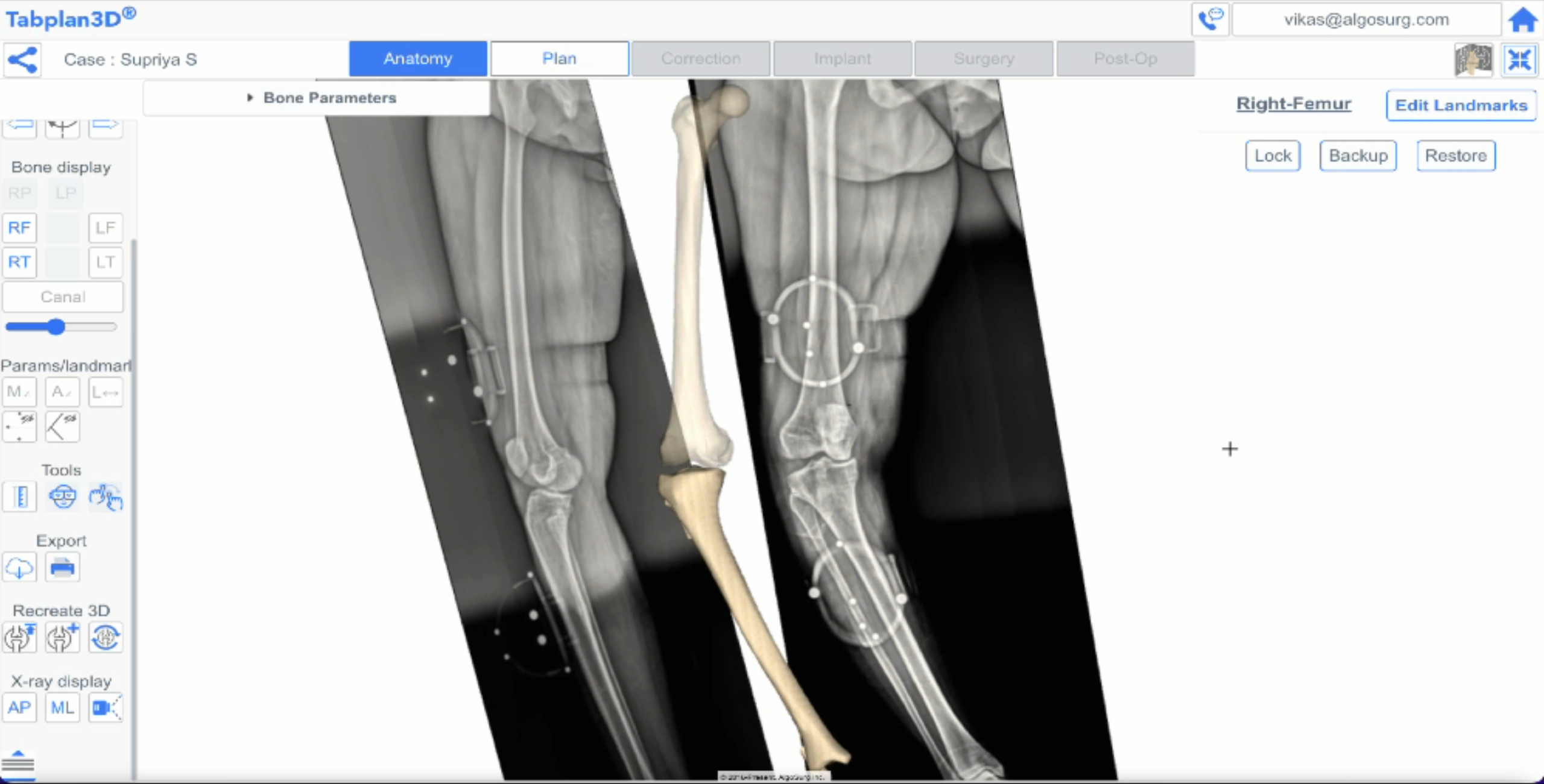
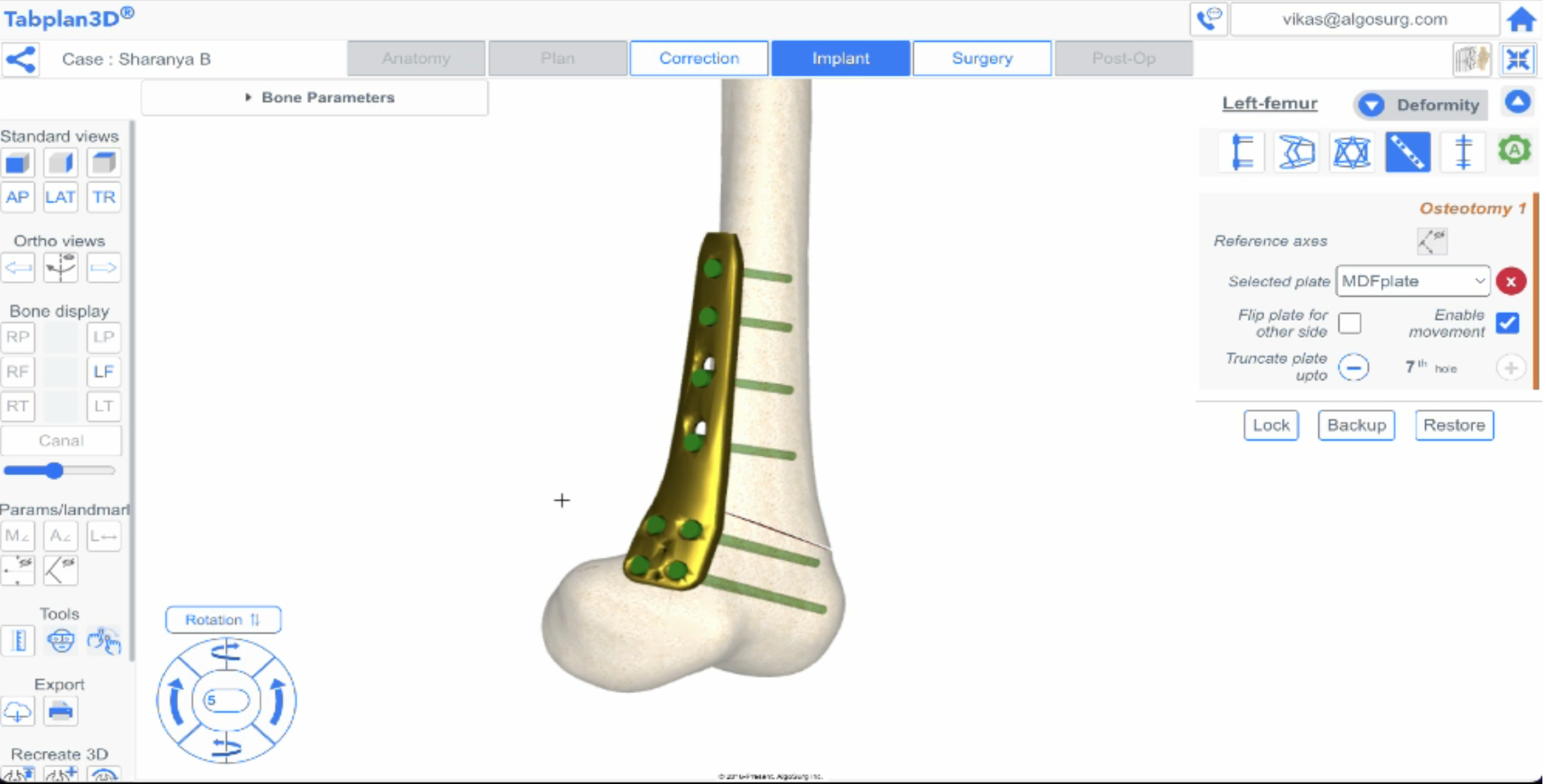
- Interactive Tools for Enhanced Surgical Planning: It provides interactive tools for patient-specific surgical planning in a 3D environment. Surgeons can manipulate the reconstructed 3D bone models, perform virtual resections, and even import 3D models of implants to plan surgeries with greater precision.
- Cost-Effectiveness: The use of readily available 2D X-ray technology reduces the need for expensive CT scans, leading to significant cost savings in diagnosis and surgical planning.
- 3D Bone Model Reconstruction: The system reconstructs 3D bone models from standard 2D X-ray images by using a pre-existing 3D bone template that is deformed to match the patient’s anatomy as depicted in the X-ray images.
- Camera Model Utilization: The Camera model includes parameters such as source-to-image distance, source-to-object distance, and image magnification to ensure accurate back-projection and 3D reconstruction.
- Novel Alignment Method: The unique alignment method accurately positions the 3D bone template in relation to the patient’s X-ray by identifying anatomical landmarks in both the 2D and 3D templates.
- Selective Anatomical Modification: Selective anatomical modification to fine-tune the template deformation based on patient-specific anatomical measurements to ensure the final 3D model accurately reflects the patient’s bone dimensions and proportions.
- Laplacian Surface Deformation (LSD): It uses LSD to create a smooth and anatomically plausible 3D bone model by deforming the template mesh while preserving its topology and shape characteristics.
- Self-Organizing Maps (SOM): To establish a precise 2D-to-2D correspondence between the projected contour of the 3D template and the actual bone contour extracted from the X-ray.
The method is designed to generate 3D bone images from 2D X-ray images. It incorporates several mechanisms: X-ray input, data import, camera model determination, contouring, bone template model input, silhouette vertices extraction, alignment, projection, selective anatomical modification, matching point, back-projecting, and template deformation. The system acquires X-ray images (at least two) of the bone, creates a model of the X-ray imaging setup, extracts the bone’s outline, and aligns a pre-existing 3D bone template with the contours. The template is then deformed using Laplacian surface deformation (LSD) to match the bone’s shape more accurately. The final output is a 3D surface model of the bone generated from the deformed template. The system assesses the accuracy of the 3D reconstruction through measurements like the mean absolute distance (MAD) between the points of the template projection contour and their corresponding closest points on the input contour. The iterative alignment process aims to minimize this MAD value, stopping when the difference in MAD between consecutive iterations falls below a pre-set threshold.
This technology has the potential to significantly impact healthcare accessibility and surgical planning. By eliminating the need for expensive and radiation-heavy CT scans, the technology could make 3D surgical planning accessible to a wider patient population, particularly in resource-limited settings where CT scanners are less available. This could lead to improved surgical accuracy, reduced complication rates, and better patient outcomes.
Its primary application domain is in healthcare, specifically for 3D pre-operative surgical planning in orthopedics and potentially other surgical specialties.