A Portable Microscope
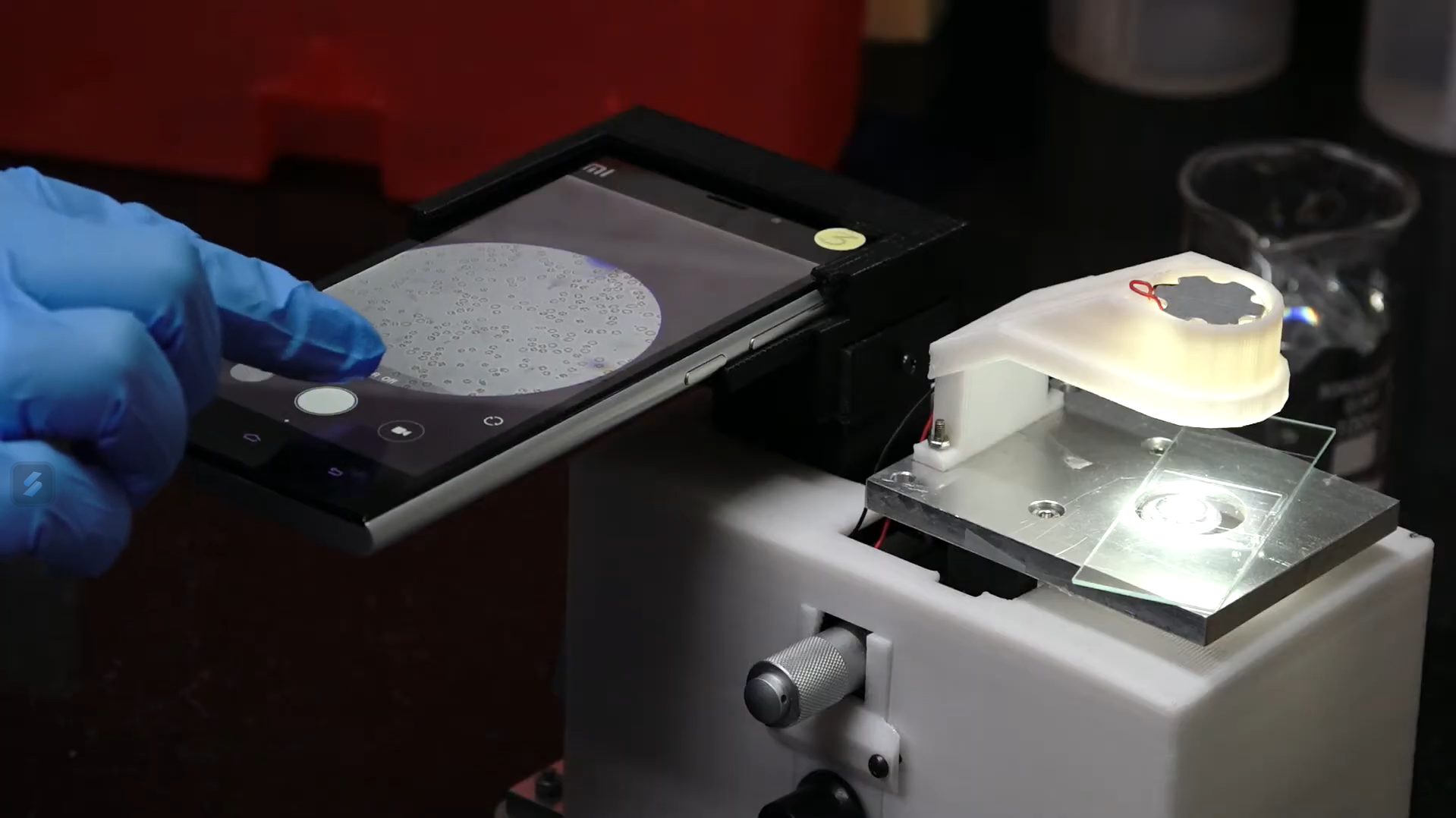
This technology describes a portable, smartphone-integrated high-magnification (2000x) imaging microscope. The compact design (8cm x 13.5cm x 12cm) incorporates two right-angular prisms to achieve a folded optical path, minimizing size while maintaining image quality. The microscope integrates seamlessly with smartphones, leveraging its camera capabilities for live imaging, image capture, recording, processing, analysis, transmission, and storage. The bench-top design ensures stability for precise focusing, surpassing the limitations of handheld portable microscopes. This innovation holds immense potential for point-of-care diagnostics, particularly in resource-limited settings, by enabling rapid on-site disease diagnosis and facilitating timely treatment.
The widespread dependence on bulky and expensive benchtop microscopes poses a significant challenge for diagnosing diseases in areas with limited resources. These traditional microscopes are not only cost-prohibitive but also hinder portability due to their size and weight. Additionally, analyzing and sharing captured images necessitates a separate computer system, further complicating the diagnostic process in remote locations. To address these limitations, there is a need for low-cost, portable microscopes with high magnification and built-in processing power, enabling direct image analysis, transmission, and, ultimately, swifter diagnosis in resource-limited settings.
- The overall dimensions of the microscope are 8 cm x 13.5 cm x 12 cm, making it highly portable compared to traditional bench-top microscopes.
- The microscope is designed to integrate with any smartphone with a camera, enabling live imaging, capturing, recording, processing, analyzing, transmitting, and storing microscopic images in real-time.
- This microscope achieves a significantly higher magnification of 2000x by combining eyepiece and objective lenses than other portable microscopes, often maxing at around 50X or 350X.
- The design employs two right-angular prisms to bend the light path by 180 degrees, reducing the overall dimensions of the microscope while maintaining a necessary optical path length to ensure high magnification and minimal aberrations.
- Unlike handheld designs, which can be unstable, this microscope is designed for bench-top use, enhancing mechanical stability and allowing for more precise focusing.
This portable single-objective microscope consists of three key components: an optical assembly for capturing magnified images, a focusing assembly for adjusting the sample position, and an illumination assembly to light up the specimen. The optical assembly is designed to achieve high magnification (up to 2000x) while maintaining image quality. The focusing assembly enables precise control over the sample stage, while the illumination assembly provides artificial light for clear image capture. The system is housed in a casing measuring only 8cm x 13.5cm x 12cm, making it portable for field use.
This portable microscope prototype offers a promising solution for improving disease diagnosis in resource-limited settings, particularly for endemic diseases like malaria and sickle cell disease that heavily impact underprivileged rural and tribal populations in regions like India and Africa. By enabling rapid on-site diagnosis, this technology has the potential to improve patient outcomes significantly. Many patients with these diseases struggle with the gap between initial, often non-confirmatory, screening tests and definitive diagnosis at distant hospitals. This delay can lead to patients dropping out of the diagnostic process altogether. The portable microscope addresses this issue by allowing for confirmed diagnosis at the point of care, facilitating the prompt initiation of disease-specific treatment. Early treatment can not only improve patient health but also potentially reduce healthcare costs associated with missed diagnoses, late diagnoses, and the development of drug resistance.
This line of digital microscopes meets the demand for high-quality indigenous inverted microscopes in various settings, including research labs (such as tissue culture microscopes) in universities and colleges, educational institutions serving large class sizes, pathology labs in tier 2/3/4 cities for basic hematology examinations and initial tests for bacterial, fungal, and parasitic infections, and IVF labs. The global microscopy market, valued at USD 6.5 billion in 2020, is projected to reach USD 8 billion by 2026.