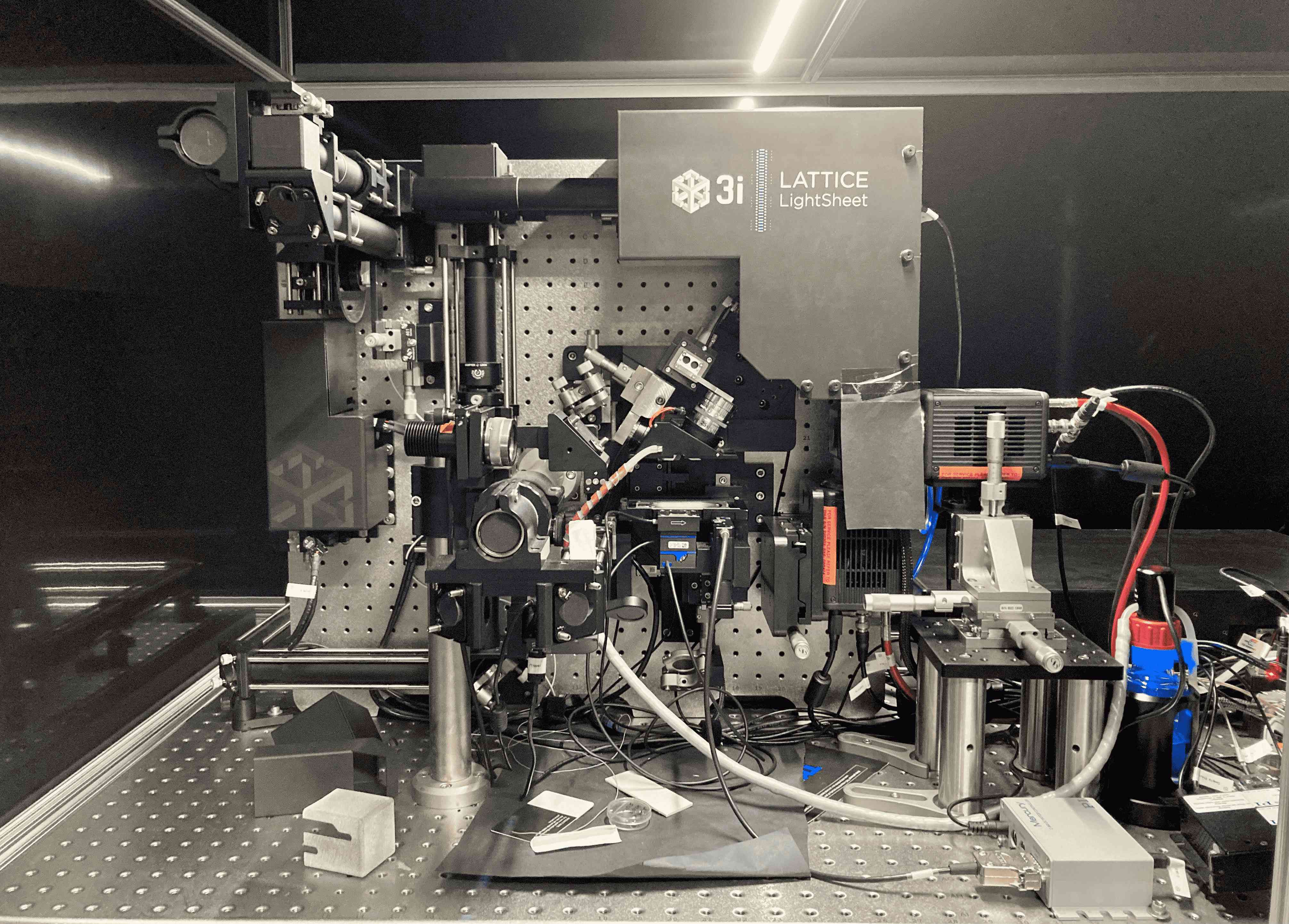
Lattice Light Sheet Microscopy Facility-IoE WCRL
Objectives: 0.71 NA 3.7 mm WD excitation objective, 1.1 NA 2 mm WD detection objective.
Magnification (effective): 62.5 X
Lasers: 488 nm, 560 nm, 642 nm
Cameras: Two Hamamatsu ORCA-Fusion BT sCMOS Camera
Incubation Stage: Temperature controller (22 – 37 o C), CO 2 , and humidity control, 8 ml and 3 ml sample chambers
Filters: 488-640T 560R custom dichroic mirror with FF01 – 446/523/600/677-25 custom made quad filter
- Semrock FF01 – 698/70
- Semrock FF01 – 510/42
- Semrock FF01 – 600/52
Sample Preparation: All samples must be prepared on a 5 mm circular coverslip. See the details later in this document.
Light Sheet Length & Thickness: Light sheet thickness is the axial resolution of a given excitation wavelength (~650 nm for 488 nm laser). Sheet length can be selected between 15 µm to 75 µm.
Data Storage: 60 TB network attached storage
Workstations: One analysis and one acquisition workstation.
Software Packages: Slidebook software (3i) for both analysis and acquisition station with Deconvolution (nearest neighbour, constrained iterative), cell/particle tracking, co-localization packages.
- Live and fixed cell imaging.
Imaging Facility, Ground Floor, Department of Biosciences & Bioengineering (BSBE), I.I.T. Bombay, Powai, Mumbai – 400076
Contact No: 022-2576-6719
- Dr. Debayan Purkait
Prof. Roop Mallik
Prof. Anirban Banerjee
Prof. Shamik Sen
Prof. Arindam Chowdhury
Prof. Nitin Kumar