- TECHNICAL SPECIFICATIONS
- SPECIAL FEATURES
- WORKING PRINCIPLE
- CENTRAL FACILITY WORKSHOP PRESENTATION
- FAQ
- PUBLICATION USING DATA FROM FACILITY
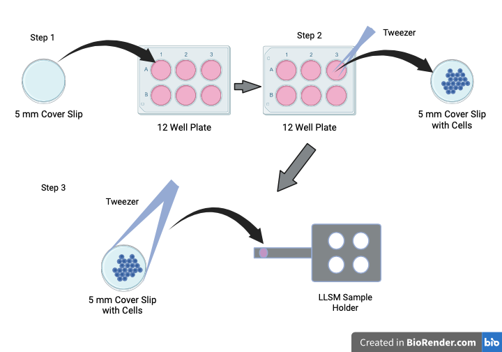
- INSTRUCTION FOR SAMPLE PREPARATION
- INSTRUCTIONS FOR USERS
- INSTRUCTIONS FOR REGESTRATION
Objectives: 0.71 NA 3.7 mm WD excitation objective, 1.1 NA 2 mm WD detection objective.
Magnification (effective): 62.5 X
Lasers: 488 nm, 560 nm, 642 nm
Cameras: Two Hamamatsu ORCA-Fusion BT sCMOS Camera
Incubation Stage: Temperature controller (22 – 37 o C), CO 2 , and humidity control, 8 ml and 3 ml sample chambers
Filters: 488-640T 560R custom dichroic mirror with FF01 – 446/523/600/677-25 custom made quad filter
- Semrock FF01 – 698/70
- Semrock FF01 – 510/42
- Semrock FF01 – 600/52
Sample Preparation: All samples must be prepared on a 5 mm circular coverslip. See the details later in this document.
Light Sheet Length & Thickness: Light sheet thickness is the axial resolution of a given excitation wavelength (~650 nm for 488 nm laser). Sheet length can be selected between 15 µm to 75 µm.
Data Storage: 60 TB network attached storage
Workstations: One analysis and one acquisition workstation.
Software Packages: Slidebook software (3i) for both analysis and acquisition station with Deconvolution (nearest neighbour, constrained iterative), cell/particle tracking, co-localization packages.
A laser beam is directed through a spatial light modulator and an annular mask to create a thin and uniform array (i.e. a lattice) of Bessel beams that illuminates the sample. This lattice is swept back-and-forth to create a thin uniform sheet of light inside the sample.
Live Cells:
Bring the cells to the imaging facility at the BSBE Dept. IIT-B one day before the scheduled imaging date. Cells must have a fluorescent marker relevant to your experiment. You must bring your own cell culture media and cell culture plates. Collect the 5 mm circular coverslips (Product Code: AGL46R5-1, Agar Scientific) from the LLSM facility. Place the coverslips inside cell culture plates and seed the cells on the coverslips (see image below). Keep cells inside cell culture incubator overnight. Incubator is available at BSBE for external users. On the day of imaging, user must provide 12 ml of culture media (at least 30 minutes prior to the imaging session) to the LLSM facility operator to calibrate the instrument.
Fixed Cells:
Collect the 5 mm circular coverslips (Product Code: AGL46R5-1, Agar Scientific) before the scheduled imaging date. Place coverslips inside cell culture plates and seed the cells having relevant fluorescent marker on the 5 mm coverslips (see image below). Fix the cells on coverslip and bring to the BSBE Dept.IIT-B.
Zebra-fish Embryo:
Early development in Zebrafish embryos can be imaged on a Light Sheet microscope. Please contact the facility manager for more information.
Inter Users: Users within IIT Bombay can register through http://drona.ircc.iitb.ac.in . The registration form should be duly filled up and all the sample details must be provided in the form. Users have to be present at the time of data acquisition and analysis by taking prior appointment with the facility in- charge. A soft copy of the duly filled registration form and the proof of payment should be emailed to the facility.
To avail the Lattice Light Sheet Microscope (LLSM) facility at IIT-B, registration is mandatory. Before the registration, the users need to discuss the experiment with the facility in-charge. Internal users are requested to discuss the experiment with the facility in-charge in-person. For the external users, a virtual meeting can be arranged by the facility in-charge, kindly send an email to facility requesting for the same.