Researchers at IIT Bombay develop a simpler and efficient method to recover immune cells grown in the lab for T-cell–based cancer therapies.
Immunotherapy, which involves boosting the body's own immune system to recognise and destroy cancer cells, has shown promising results in cancer treatment. In immunotherapies such as CAR T-cell, doctors take T-cells (a type of immune cell) from a patient’s blood and modify them in the laboratory so they can better recognise and attack cancer cells. These modified cells are then grown in large numbers and infused back into the patient’s bloodstream to help fight cancer.
A key requirement for T-cell-based immunotherapy is an ample supply of healthy, active T- cells. These cells, grown outside the body, must be collected gently so that they remain alive and functional when returned to the patient. Finding safe and efficient ways to grow T-cells and retrieve them is therefore an important part of making these therapies work.
In a recent study, a research team from the Department of Biosciences and Bioengineering at the Indian Institute of Technology Bombay (IIT Bombay), led by Prof. Prakriti Tayalia, has developed a simple method to gently recover T-cells after growing them in the laboratory. The study was carried out in collaboration with Prof. Neil Cameron of Monash University and published in the journal Biomaterials Science.
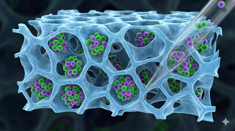
In many laboratories, T cells are grown on flat plastic dishes. While easy to use, these flat surfaces do not reflect how cells behave inside the human body. In the body, T-cells move through complex tissue spaces lined with densely packed cells and three-dimensional networks made of fine fibres. To better mimic this natural environment, researchers have begun using three-dimensional scaffolds, which may provide a porous as well as fibre-like structure for cells to grow in.
Prof. Tayalia’s team works with a specific type of scaffold made using a process called electrospinning. These electrospun scaffolds look like thin mats made of very fine fibres, similar to a dense fishing net. Earlier studies by the team and other research groups have shown that T-cells grown on such scaffolds become more active and multiply faster.
However, these advantages come with a challenge. As T-cells move deep into the spaces between the fibres, they become difficult to remove. For any therapy, cells must be collected, tested, and finally delivered to patients. If too many cells remain trapped in the scaffold, the process becomes inefficient.
“Cell recovery sounds simple on paper, but in practice it turns out to be one of the biggest challenges,” Prof. Tayalia explains. “Without enough healthy cells, you cannot test them properly or use them for therapy.
”To address this problem, the team grew Jurkat T-cells (a human cell line grown and used in the laboratory to study T-cell biology, cancer and HIV) inside electrospun scaffolds made from a material called polycaprolactone. Under a microscope, the researchers observed that the cells actively moved into the scaffold and became tightly lodged between the fibres. Even strong flushing with a pipette using the growth medium could not remove all the cells, especially those stuck at fibre junctions.
“Theoretically, T-cells are considered easy to handle because they are ‘suspension cells’— they usually float freely in liquid. In reality, when placed inside a dense fibre network, they grip tightly,” says Dr. Jaydeep Das, the study’s first author.
The researchers then tested three different methods for collecting the cells. The first was simple manual flushing in the growth medium using a pipette. The second method used TrypLE—a version of the enzyme trypsin that helps detach cells in laboratories. The third method used Accutase, a milder enzyme designed to remove cells more gently.
For each method, the researchers measured three outcomes: The number of cells that were recovered, the number that remained alive, and whether the cells could continue to grow afterwards. While the total cell recovery was comparable in all three methods, the accutase- based removal yielded more viable cells. To test how well the recovered cells functioned, the team allowed them to grow for a week after recovery. “We didn’t just want living cells. We wanted cells that still knew how to behave like T-cells,” says Dr. Das.
Cells recovered using trypsin showed higher cell death. Some of the surviving cells also lost important behaviours needed for proper immune function. In contrast, cells recovered with accutase survived in greater numbers and behaved more like healthy T-cells. They formed clusters, an essential step before T-cells divide, and continued to grow well after recovery.
“Harsh treatments to cells, using enzymes such as trypsin, can damage key surface proteins needed for immune signalling and activation, reducing the cell’s therapeutic usefulness. Accutase appears mild enough to avoid this problem,” says Prof. Tayalia.
The study's findings could help laboratories use such scaffolds when preparing cells for therapies such as CAR T-cell treatment. “If we want these advanced therapies to reach patients, every step matters. How we grow cells, and how we retrieve them, can make a real difference,” notes Prof. Tayalia.
Building on this work, the team has also found that T-cells grown on scaffolds can kill cancer cells more effectively. In the future, they plan to test these findings in animal models and explore the possibility of placing T-cell–loaded scaffolds directly inside the body.
Funding information:
Department of Biotechnology, Monash University, Department of Health Research—Indian Council of Medical Research, Australian Research Council, The European Union.
Prof. Prakriti Tayalia, Department of Biosciences and Bioengineering, Indian Institute of Technology Bombay, Mumbai