Cryo high resolution transmission electron microscope facility

The facility consists of following equipment.
- Aberration corrected Cryo-HRTEM (Jeol NEOARM 200F)
- Cryo-HRTEM (Jeol JEM 2100)
- Cryo-Ultramicrotome (Leica EM UC7)
- Freeze fracture (Leica EM ACE900)
- Cryo-plunger (Leica EM GP2)
1.Aberration corrected Cryo-HRTEM
"NEOARM" / JEM-ARM200F comes with unique cold field emission gun (Cold FEG) and a new Cs corrector (ASCOR) that compensates for higher order aberrations. The combination of a Cold FEG and ASCOR enables atomic-resolution imaging at not only 200 kV accelerating voltage, but also a low voltage of 30 kV.
"NEOARM" is also equipped with an automated aberration correction system that incorporates aberration correction algorithm for automatic fast and precise aberration correction. This system enables higher-throughput atomic-resolution imaging.
Furthermore, a new STEM detector that provides enhanced contrast of light elements is incorporated as a standard unit. Contrast enhancement of light elements is achieved by a new STEM imaging technique, facilitating observation of light-element materials, even at low accelerating voltages.
Make and Model: JEOL NEOARM 200F
Date of Installation: 19th March 2024
Specifications:
- Point resolution: 0.27 nm
- Lattice resolution: 0.14 nm
- STEM resolution: 0.082 nm
- Electron Source: Cold field emission gun.
- Accelerating Voltage: 80 kV & 200 kV
- Magnification: X2000 - X1500000
- Specimen Tilt angle X/Y: +- 35°
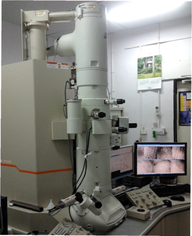
2.Cryo-HRTEM
The JEM-2100 is a multipurpose, 200 kV analytical electron microscope with LaB6 source. supports research and development in wide scientific fields, for biology to materials researches.
Make and Model: : JEOL JEM 2100
Date of Installation: 12th Aug 2010
Specifications:
- Point resolution: 0.194 nm
- lattice resolution: 0.14 nm.
- Max. Eucentric tilt: ±80°
- Magnification: 50x to 1500000x
- Electron Source: LaB6 emitter
- Accelerating Voltage: 80,120,200 kV
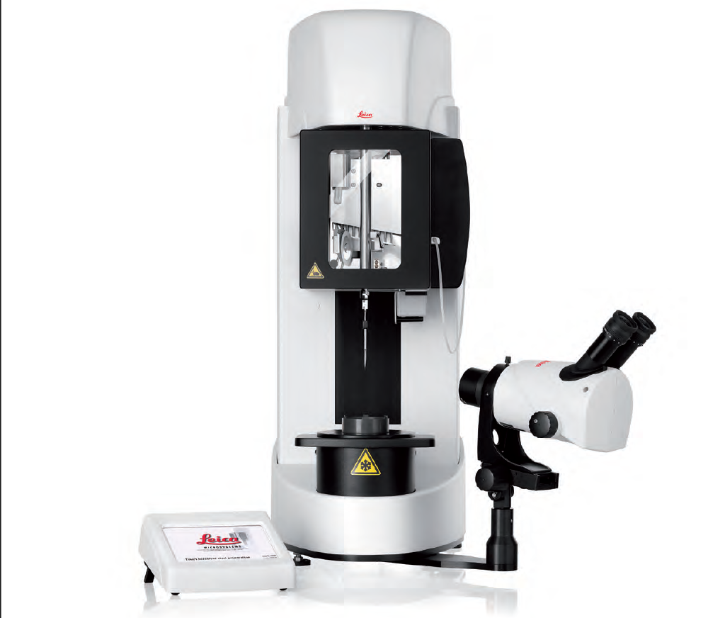
3.Cryo-Ultramicrotome
Make and Model: : Leica EM UC7
Useful to prepare high-quality ultra- or semi-thin sections for your transmission electron or light microscope investigation whilst simultaneously creating perfectly smooth block face surfaces for atomic force, scanning electron, or incident light microscopy. For ultrathin cryo- sections or surfacing of cryogenic material, EM UC7 ultramicrotome can be equipped with the EM FC7 low-temperature sectioning system as needed.
4.Freeze fracture
Make and Model: : Leica EM ACE900
Freeze fracture describes the technique of breaking a frozen specimen to reveal internal structures. Freeze etching is the sublimation of surface ice under vacuum to reveal details of the fractured face that were originally hidden. A metal/carbon mix enables the sample to be imaged in a SEM (blockface) or TEM (replica).
5.Cryo-plunger
Make and Model: : Leica EM GP2
System offers automated vitrification to provide fast, easy, and reproducible sample preparation for cryo-EM. It performs the cryo-fixation process at constant physical and mechanical conditions like temperature, relative humidity, blotting conditions, and freezing velocity. This ensures high-quality cryo-fixation results and a high sample preparation throughput prior to cryo-TEM observation.
- TEM/HR Imaging & Diffraction
- CRYO Imaging
- STEM Imaging
- EDS analysis (spectrum+ Image)
- EDS Line Scan analysis
- EDS Mapping analysis
- Typical Samples: powder samples, CNT, Nano particles, Ferrite and Plant extracts etc.
- Suspension of nanoparticles including vesicles, liposomes, solid lipid nanoparticles, cores shell particles,hard and soft latex, nano medicines across all systems of medicines, biological cells and organelles, tissue blocks and similar samples.
- Staining: Positive staining, Negative Staining and Fume hood staining
- . Embedding & sectioning: The specimen is placed in a mold filled with liquid resin and cured into a hard block using heat. After this, specimens embedded in hardened resin can be sectioned extremely thin, at less than 100 nm.
- Freeze facture: Breaking a frozen specimen to reveal internal structures. Samples to be imaged in a SEM (block-face) or TEM (replica)
- Cryo-Plunging: Samples are cooled so rapidly that the surrounding water molecules do not have time to crystallize.
- Dispersion: Powder samples will be dispersed in the solvent and after ultrasonication, it will be loaded/drop casted on the TEM grid. The grid will be dried under the IR lamp. Dispersion will be done by ultrasonication.
- Nanotechnology, Biology and Life sciences, Material science, Pharmaceutical analysis, Semiconductors.
- Prof. Jayesh Bellare
Cryo-HRTEM Lab, Room No.102A, Ground floor, Silicate Lab, Chemical Engineering Department. IIT Bombay, Mumbai - 400 076.
Contact No: 022-2159-6166
Mr. Ashish Karale
Project Manager