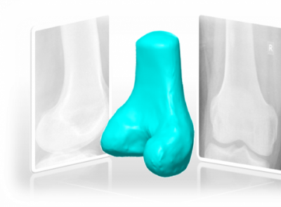
“Can we get a 3D model from 2D X-ray images of a bone from a patient’s body?” asked a top surgeon from Hinduja hospital, who wanted an alternative to the current practice. The OrthoCAD lab’s software can do this within a minute, with atmost two X-ray images.
The challenge looked impossible, because as any engineer knows, 2D views can be generated from a 3D model but not vice versa. But Dr. Manish Agarwal, the surgeon, quipped that if he can reconstruct the model in his mind, then why can’t we develop a computer program to do the same. The idea was to generate 3D model using X-ray images unlike reconstructing a 3D model from CT scanning which involved several hundred times more radiation, besides being more expensive to the patient.
Thus started the journey of Vikas Karade a research scholar and Professor B. Ravi at OrthoCAD Lab, to find a way to solve the above problem. Aptly named ‘XrayTo3D’, their program can convert 2D Xray images of a bone (femur, in this case) from a patient into a 3D model. A demo version exists on the OrthoCAD lab website, which can be freely used by any clinician. This work fetched Vikas the ‘Gandhian Young Technological Innovation Award’ on 2014.
The input is either one or two X-ray images taken at perpendicular planes and a 3D template of the corresponding bone. The template needs to be created only once (for each bone) from CT images of a healthy patient. The template is projected on the two planes of the X-ray images. The projections are matched in shape with the X-ray images by the algorithm and the modified projections are converted back to a 3D model. It takes less than a minute to generate the 3D shape of the patient’s bone, with an error of less than 1.5 mm compared to that reconstructed from CT images.
The program can be used by orthopedic surgeons to plan surgeries in 3D without using radiation-intensive and expensive CT scans, which is a boon even in rural areas where CT scan machines are rare. Vikas is now implementing the program for use on tablets and smartphones, making it orthopedic surgeons’ next best friend and ben
“Can we get a 3D model from 2D X-ray images of a bone from a patient’s body?” asked a top surgeon from Hinduja hospital, who wanted an alternative to the current practice. The OrthoCAD lab’s software can do this within a minute, with atmost two X-ray images.
The challenge looked impossible, because as any engineer knows, 2D views can be generated from a 3D model but not vice versa. But Dr. Manish Agarwal, the surgeon, quipped that if he can reconstruct the model in his mind, then why can’t we develop a computer program to do the same. The idea was to generate 3D model using X-ray images unlike reconstructing a 3D model from CT scanning which involved several hundred times more radiation, besides being more expensive to the patient.
Thus started the journey of Vikas Karade a research scholar and Professor B. Ravi at OrthoCAD Lab, to find a way to solve the above problem. Aptly named ‘XrayTo3D’, their program can convert 2D Xray images of a bone (femur, in this case) from a patient into a 3D model. A demo version exists on the OrthoCAD lab website, which can be freely used by any clinician. This work fetched Vikas the ‘Gandhian Young Technological Innovation Award’ on 2014.
The input is either one or two X-ray images taken at perpendicular planes and a 3D template of the corresponding bone. The template needs to be created only once (for each bone) from CT images of a healthy patient. The template is projected on the two planes of the X-ray images. The projections are matched in shape with the X-ray images by the algorithm and the modified projections are converted back to a 3D model. It takes less than a minute to generate the 3D shape of the patient’s bone, with an error of less than 1.5 mm compared to that reconstructed from CT images.
The program can be used by orthopedic surgeons to plan surgeries in 3D without using radiation-intensive and expensive CT scans, which is a boon even in rural areas where CT scan machines are rare. Vikas is now implementing the program for use on tablets and smartphones, making it orthopedic surgeons’ next best friend and benefitting thousands of patients.
Prof. B Ravi