- TECHNICAL SPECIFICATIONS
- SPECIAL FEATURES
- WORKING PRINCIPLE
- CENTRAL FACILITY WORKSHOP PRESENTATION
- FAQ
- PUBLICATION USING DATA FROM FACILITY
- INSTRUCTION FOR SAMPLE PREPARATION
- INSTRUCTIONS FOR USERS
- INSTRUCTIONS FOR REGESTRATION
Microscope: Fully motorized and computer-controlled Nikon ECLIPSE Ti2 microscope (inverted) with motorized Peizo Z-axis focus drive optimal for super-resolution imaging with resolution/step size of 2.5 nm.
Objectives: PLAN APO λD 10X (air), Plan Apo λ 40x(air) Ph2 DM/0.95 NA, PLAN APO λD 60x Oil/1.40 NA, SR HP Apo TIRF 100XAC Oil/1.49 NA.
Confocal Scanning Module: Up to 0.001 frames/sec scanning speed at 8192 x 8192 pixels. Four channels in a proprietary sensitive GaAsP spectral detector can be simultaneously captured. One transmitted light PMT detector for laser scanning DIC imaging is available.
Confocal Lasers: All DPSS/Diode high power, long life lasers and pre-aligned having AOTF control with Solid state (405 nm, 445 nm, 488 nm, 561 nm, 647/640/635 nm, 514 nm) Confocal usage.
STORM Lasers: High-power solid-state lasers 405 nm, 488 nm, 561 nm, and 640 nm for STORM imaging.
VT i-SIM Lasers: High-Speed 4-Position Emission Filter Changer. Excitation: Four solid-state lasers selectable from within the visible range (405 nm, 488 nm, 561 nm, 642 nm).
Filters: Four fixed filters (DAPI, FITC, TRITC, CFP) and one Quad band filter are available.
Software: NIS-elements acquisition software from Nikon with 3D, ROI, FRAP, FRET, N-STORM, C-STORM, VT-iSIM, and stitching modules are provided.
- On-stage incubation system with temperature and CO2 control for long-time live cell imaging.
- The incubation system can also control N2/O2 for hypoxia experiments.
- The system is also equipped with Perfect Focus System (PFS), a hardware LED-based drift compensator, for long-time live cell imaging.
1. VT-iSIM Structured Illumination Microscopy:
The leading ISM technique for live cell imaging is Instant SIM and in particular, the VT-iSIM, which allows for super-resolution fluorescence imaging to be achieved at high frame rates with reduced photobleaching. The iSIM utilises a galvanometer scanner to scan a 2-Dimensional (2D) array of points at the sample plane. The 2D-array scan architecture was developed by VisiTech in the early 2000’s and became the basis for the analogue based ISM techniques as per York, Shroff, et al (doi:10.1038/nmeth.2687). For the iSIM, it means that the emission signal is de-scanned by the galvo. Hence, you have a conjugate image plane in an isolated emission path where you can place the re-assignment optic (a μLens array) and then re-scan the emission onto the camera, as per figure.
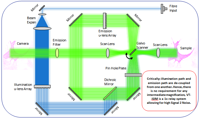
The re-assignment optic shrinks each individual emission Point Spread Function (PSF) in the array by 0.5x. This subsequently doubles the Numerical Aperture (NA) of the light in the emission path. In the iSIM; as the re-assignment optic is in a conjugate image plane in the emission path (and not in the excitation path), there is no requirement for intermediary magnification and hence much improved S2N.
The re-assignment optic is a μLens array. μLens are designed to collect as much light as possible. If the out-of-focus light is not removed first, it will be collected by the re-assignment optic and focused through the pin-holes, thus increasing the overall background signal. Obviously, as the iSIM has the re-assignment optic in a conjugate image plane which is between the pinhole array and the camera, the out-of-focus light is removed before the re-assignment. This results in much lower levels of background/noise.
1. N-STORM (Nikon):
STochastic Optical Reconstruction Microscopy (STORM) reconstructs a super-resolution image by combining the high-accuracy localization information of individual fluorophores in three dimensions and multiple colors
N-STORM uses stochastic activation of relatively small numbers of fluorophores using very low-intensity light. This random stochastic "activation" of fluorophores allows temporal separation of individual molecules, enabling high-precision Gaussian fitting of each fluorophore image in XY. By utilizing special 3D-STORM optics, N-STORM can also localize individual molecules along the Z-axis with high precision. Computationally combining molecular coordinates in three dimensions results in super-resolution 3D images.

High-precision Z-axis position detection: Using a cylindrical lens that asymmetrically condenses light beams in either X or Y direction, Z-axis molecule locations can be determined with an accuracy of about 50 nm. Location in Z is determined by detecting the orientation of the astigmatism-induced stretch in the X or Y direction and the size of the out-of-focus point images. 3D fluorescent images can be reconstructed by combining the determined Z-axis location information with XY-axis location information.
This FAQ deals with the operational aspects of the facility. If you would like to suggest a question, do feel free to drop an email to <srconfocal@gmail.com>
A. I need to do simple slide imaging. Which confocal modality should I use?
You could use either system as long as you are imaging up to four fluorophores (Dapi, Green, Red, and Far-red). If you want to capture images in confocal mode, you have to use the laser scanning confocal microscope. If you want to image fluorophores for high resolution, you need to use either i-SIM or STORM modality for your sample.
B. What consumable items should I bring with me? What items will be provided at the facility?
The facility will only provide the immersion oil for the objectives and the lens cleaning tissues. Everything else that you may need during imaging (e.g. gloves, pipettes, tips, regular tissue rolls, aluminium foil to cover sample, etc.) you will have to bring yourself. If you are in doubt, please speak to the operators in advance.
C. I need to use the SR confocal microscope. Do I need to train as a TA?
If your usage is infrequent (less than twice per month), the operators or the existing TAs can do the imaging for you. If your research project heavily depends on the use of the SR confocal facility, it would be better if you trained as a TA. Do remember, training as a TA comes with certain duties, such as imaging other people’s samples.
D. What does being a TA involve?
The job of a TA is to help us run the facility smoothly and image other people’s samples. You will have a do a minimum of 6 hours of TA duty per week just like the TAs allotted to other central facility equipment. This is non-negotiable. If you are a first-year PhD student with loads of coursework, we suggest that you come back after a year. The upside is that you will get really proficient in using a state-of-the-art SR confocal microscope. You will also be able to book slots during ‘off’ hours (between 6 pm and 9 am) to run samples for yourself or your research group. On the whole, it should be a very useful learning experience for you.
E. I think I need to train as a TA. What should I do?
The first thing you should do is to check with your advisor on whether both of you agree with the time commitment. If you are a non-BSBE student, you need to contact the TA coordinator of your department to see if you could be assigned as a TA in the central facility. If the answer to both questions is ‘yes’, send an email to the convener of the microscope where you want to train. We will take over from there.
F. I booked a slot, but my sample is not ready. What should I do?
This can happen once in a while, so don’t worry. Send an email to <srconfocal@gmail.com> and call the operator on his mobile phone as soon as you realize that you cannot make it to your slot. This is a matter of courtesy to ensure that other people can use your slot. If this happens too many times, clearly you are not planning your experiments very well and we will take a strict view of it.
G. I need to do live cell imaging. Which microscope should I go for?
If you are imaging swimming bacteria, sperm cells, etc. you should definitely choose the spinning disc system. If your live cell imaging involves four fluorophores, you have to choose the i-SIM SR confocal.
H. I need to book more than one consecutive slot. Can I get it?
Sure, if you can justify why. We need to be fair to every user while assigning slots. We will give you as many slots as you need to image all your samples, but they will be distributed over several days.
I. I don’t have any fluorophore in my sample. Can I still use the confocal system?
Just remember that it won’t be SR confocal imaging, i.e. you won’t be blocking the out-of-plane light. In such a case, the Perfect Focus System (PFS) will be very useful to you to ensure that at least one of the Z-stack images remains in focus throughout.
J. Can I request a particular TA to image my sample?
No. All TAs have done the same training and it should not matter who images your sample. The conveners have framed this policy to ensure that no single lab/TA monopolizes the use of the facility. If you have any apprehensions about any TA, feel free bring it to the notice of the convener immediately.
- To achieve the best results during examination in the Super-Resolution Confocal Microscope (SRM), perfect SRM sample preparation (for i-SIM and STORM) is required.
- The required techniques depend on the type of samples (biological samples, material samples) as well as on the application.
- Only if each step of sample preparation is of the highest quality, one can get optimum results from a high-resolution STORM microscope.
- Currently, we can image fixed samples sealed between a glass slide and a cover slip. Do not bring samples without sealing them with a cover slip.
- 35 mm diameter Petri dishes. Please use specially available imaging Petri dishes with coverslip bottoms if you wish to use oil immersion objectives.
- Multi-well plate dishes. Please use a specially available imaging multi-well plate with coverslip bottoms if you wish to use oil immersion objectives.
- For the STORM sample preparation, please find the link below. https://drive.google.com/drive/folders/1ECHlv0TUvUzwLi50cE0RR97LlqIpuUR-?usp=sharing
- Users should know what kind of sample preparation is required for his/her samples.
- Please mention what fluorophores you have used in your sample (excitation/emission spectra) when you make a request.
- Users must be available throughout the imaging.
- Only online registration through the IRCC webpage will be accepted. If you need to cancel your slot, send an email immediately with an explanation.
- Slots will be provided on a first-come first-served basis.
- The slots are from 9 am - 11 am, 11 am - 1 pm, 2 pm - 4 pm, 4 pm - 6 pm. You can request two consecutive slots only once a week. If your experiment needs more time (e.g. A long time live cell imaging, etc.), please drop an email to srconfocal@gmail.com or pradips@iitb.ac.in and CC Prof. Santanu Ghosh santanughosh@iitb.ac.in so that we can deal with your specific requirement.
- The non-office hours slots are of 3 hours and it starts from 6 pm to the next day 9 am. (6 pm - 9 pm, 9 pm - 12 am, 12 am - 3 am, 3 am - 6 am, and 6 am - 9 am)
- USB drives are strictly not allowed for copying data to minimize virus-related issues. The data can be shared in the cloud or you need to bring a new blank CD/DVD to transfer your data. All data must be transferred within 7 days of imaging. Without exception.
- Register online through the IRCC webpage.
Link- https://drona.ircc.iitb.ac.in/ircc/NewFac/CentralFacilities.jsp
- After the slot booking request is accepted, please contact the operator (Mr. Pradip Shinde 022-2159 6719/6746) to discuss the details of your experiment.