
IIT Bombay researchers develop a low-cost point-of-care device that can screen for anaemia and sickle cell disease using a drop of blood.
Blood smear tests are routinely conducted in pathology laboratories to screen for several diseases and conditions. However, remote places and smaller hospitals still lack access to well-equipped testing facilities, even for common health issues like anaemia, in which haemoglobin stays low and leaves the body short of oxygen. Another disease of the red blood cells, sickle cell disease (SCD; a genetic disorder that makes red blood cells rigid and prone to blocking small vessels), is prevalent in India’s tribal communities, but often remains neglected.
A research team from the Department of Chemical Engineering at the Indian Institute of Technology Bombay (IIT Bombay), led by Prof. Mahesh Tirumkudulu, has developed a simple, low-cost point-of-care approach that uses the length of a blood smear (the distance that a drop of blood spreads across a glass slide) to detect anaemia, and detect and monitor treatment for sickle cell disease. Dr. Mahrukh Mir, a former PhD scholar in Prof. Tirumkudulu's lab, is the first author and Bhavesh Raicha of Valsad Raktdan Kendra is a co-author of this study, published in the journal Lab on a Chip.
In pathology labs, a blood smear is often examined under a microscope to check cell shape, count, or parasites like malaria. A technician creates a blood smear by placing a drop of blood on a slide and dragging a second slide across it to make a thin film. Though one of the oldest and most routine procedures in medicine, manual blood smears vary in size and thickness depending on the person performing the procedure.
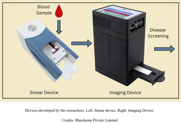
Prof. Tirumkudulu’s team built their own automated device to have a uniform method of creating a smear. The device holds two glass slides at a fixed angle to each other. A drop of blood is placed on the lower slide, and the upper slide is brought into contact with it and then moves off at a set, controlled speed. As the blood spreads on the lower slide, its film becomes thinner until it ends, creating a measurable smear length.
Under these controlled conditions, the researchers observed that blood from different people gives different smear lengths, and this difference holds useful information.
In their previous study, the researchers showed mathematically that a smear could capture blood viscosity. Prof. Tirumkudulu explains, “More viscous blood spreads less easily and therefore produces a shorter and thicker smear under the same conditions.” They confirmed their results with conventional rheometer-based (an instrument that measures how liquids flow) measurement techniques.
Once the team confirmed that the method could sense small differences in blood viscosity, they decided to investigate whether those differences could help identify disease.
Blood viscosity depends on haematocrit (the proportion of blood made up of red blood cells), plasma viscosity, and how easily red blood cells change shape. These change with disease: anaemia lowers red blood cell concentration and haemoglobin, while sickle cell disease makes red blood cells rigid.
To explore screening for severe anaemia, the researchers studied 516 individuals and compared their blood viscosity with haemoglobin measurements. The data showed that viscosity increased with haemoglobin concentration, and the researchers identified approximate viscosity thresholds associated with low haemoglobin corresponding to severe anaemia (haemoglobin below 8 g/dL). Results showed that 95.1% of people with the condition and 86% of people without the condition were correctly identified, with a haemoglobin accuracy of about ±0.8 g/dL.
“A viscosity value by itself doesn't have diagnostic value,” Prof. Tirumkudulu notes. “Tell a doctor a patient's blood viscosity value, and they can't do much with that. Tell them the haemoglobin is eight, and they know exactly what to do.”
The researchers also developed another approach based on imaging that estimates haemoglobin from the colour intensity of the dried blood film rather than its length alone. Tested on more than 1,000 clinical patients, this method showed close agreement with standard haemoglobin measurements, with an accuracy of about ±0.5 g/dL, and could detect haemoglobin levels as low as 2.0 g/dL.
Screening for sickle cell disease (SCD), however, requires a different procedure. People with SCD produce an altered form of haemoglobin (HbS). A sample containing HbS doesn’t necessarily show a large change in viscosity under normal oxygen conditions in the blood, but blood tends to become more viscous when blood oxygen is low. Thus, smear length alone cannot detect SCD. Instead of mere smear length, the researchers used the difference between the smear length of the blood sample used directly and the smear length of the deoxygenated blood sample.
The researchers used sodium metabisulfite, an inexpensive and easily available chemical, to deoxygenate the blood samples (sickling treatment). Comparing the two smear lengths indicates how viscous the blood has become after deoxygenation, which can be mapped to the percentage increase in viscosity.
“In the case of a person without SCD, there is hardly any change (in viscosity of the blood after removing oxygen),” says Dr. Mahrukh. If a person has the sickle gene from one parent (trait), viscosity increases slightly, whereas if a person has the sickle gene from both parents (full SCD), this increase is, on average, 125%.
Field tests of the method on 394 people in the tribal belt of Maharashtra and central India confirmed the effectiveness of the researchers’ approach. In healthy people, blood viscosity rose by less than 17% after deoxygenation. In people carrying the sickle cell trait, it increased by 17 to 70%. In people with full SCD, it increased by more than 70%. This clear distinction allowed the field test to identify the disease correctly in about 95% cases, in both identifying true cases and correctly clearing healthy people.
The test also worked on patients taking hydroxyurea, the primary drug used to treat SCD. Hydroxyurea helps only about half of patients, and treatment response can vary. In these patients, the viscosity rise dropped from around 125% to about 70%, showing that the drug was working and that the test can be used to monitor the treatment.
The simplicity of their method makes it attractive, according to the researchers. They report that their patented automated smearing device can be manufactured cost-effectively. Also, the test itself requires only a pipette, glass slides, an inexpensive chemical, vials for the sickling treatment, and a ruler or imaging device to measure smear length.
“This makes our technique particularly useful in places where conventional diagnostic laboratories are difficult to access, and it doesn't require any special expertise to use,” says Dr. Mahrukh.
The researchers point out that the blood viscosity cutoffs vary for various groups of people because certain genetic differences affect how blood reacts when oxygen levels drop. Also, the anaemia test could miss identifying some patients when the usual link between viscosity and anaemia does not hold, such as in a rare condition called hyperviscosity syndrome, where blood plasma is unusually thick.
Currently, the technology is licensed to Rheoheme Private Limited, an IIT Bombay startup. The team is now working with the National Institute for Research in Tribal Health, Jabalpur, and All India Institute of Medical Sciences (AIIMS) Nagpur, toward regulatory approval, while exploring extending the device to measure clotting time and glucose levels.
“Our initial aim was to understand the physics of spreading blood drop, and that led to the medical applications,” Prof. Tirumkudulu concludes while reflecting on their journey from a fundamental study of fluid mechanics to a possible diagnostic tool.
Prof. Mahesh S. Tirumkudulu, Department of Chemical Engineering, Indian Institute of Technology, Bombay, Powai